Trivehexin PET/CT Scan
A highly sensitive imaging technique that uses the Trivehexin tracer to accurately
detect parathyroid adenomas by targeting specific molecular markers present in
abnormal parathyroid tissue.
About Trivehexin PET/CT Scan
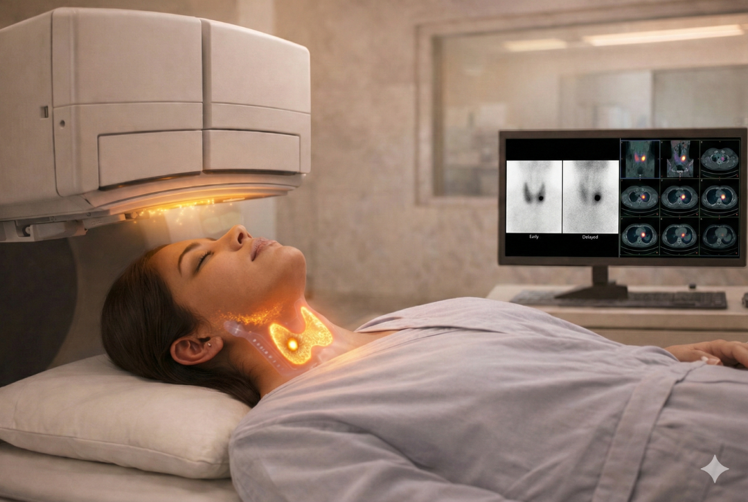
A Trivehexin PET/CT scan is an advanced molecular imaging technique used to detect and localize parathyroid adenomas, the most common cause of primary hyperparathyroidism. It combines Positron Emission Tomography (PET) and Computed Tomography (CT) to produce detailed functional and anatomical images. After injection, the rivehexin tracer binds to abnormal parathyroid tissue. PET identifies areas of increased activity, while CT provides precise structural detail. Together, they enable accurate localization, sizing, and assessment of adenomas, improving diagnosis and surgical planning.
Uses of Trivehexin PET/CT Scan
Trivehexin PET/CT scans are particularly helpful in diagnosing and managing parathyroid adenomas and hyperparathyroidism. Below are some key uses of this scan in clinical practice:
01. Detecting Parathyroid Adenomas
The scan is highly sensitive in identifying parathyroid adenomas, which are small
benign tumors responsible for excess parathyroid hormone (PTH) production.
02. Precise Localization of Abnormal Glands
Trivehexin PET/CT helps accurately identify the exact location of abnormal
parathyroid glands, even when they are very small or located in unusual positions.
03. Detection of Ectopic Parathyroid Tissue
Sometimes parathyroid glands can be located in unusual areas such as the chest or mediastinum. This scan helps detect such ectopic glands with high accuracy.
04. Planning Minimally Invasive Surgery
Precise localization allows surgeons to perform targeted minimally invasive parathyroid surgery, reducing operation time and improving outcomes.
05. Detecting Recurrent or Persistent Disease
In patients who have undergone previous surgery but still have high PTH levels, this scan helps identify residual or recurrent parathyroid adenomas.
06. Problem-Solving When Other Scans Are Negative
Trivehexin PET/CT is especially useful when conventional imaging methods such as ultrasound or sestamibi scans fail to clearly locate the abnormal gland.
Procedure for Trivehexin PET/CT Scan
-
Injection of Trivehexin Tracer
A small amount of the Trivehexin tracer is injected into a vein. This tracer circulates through the bloodstream and attaches to abnormal parathyroid tissue.
-
Waiting Period
After the injection, you will be asked to wait for about 45 minutes. During this time, the tracer accumulates in the parathyroid adenoma.
-
Scanning Process
The scan includes both PET and CT imaging, performed in a single session. The PET scan detects tracer activity, while the CT scan provides detailed anatomical images of the neck and chest. The scanning process usually takes around 10–15 minutes.
-
Comfortable and Non-Invasive Procedure
FDG PET/CT scans help identify areas of infection or inflammation in the body, aiding in the diagnosis and treatment of various conditions.
-
Comfortable and Non-Invasive Procedure
The scan is safe, painless, and non-invasive. Patients simply lie still on the scanner table while images are acquired.
Benefits of Trivehexin PET/CT Scan
-
Highly Sensitive Detection
Trivehexin PET/CT can detect very small parathyroid adenomas, even when conventional imaging tests fail.
-
Precise Localization
The scan provides accurate information about the exact location of abnormal glands, helping surgeons plan targeted treatment.
-
Better Surgical Outcomes
Accurate imaging allows surgeons to perform minimally invasive surgery, improving success rates and reducing complications.
-
Useful in Complex Cases
This scan is particularly valuable in patients with recurrent disease, prior neck surgery, or inconclusive previous imaging studies.
Subsribe To Our Newsletter
Stay in touch with us to get latest news and special offers.