Advanced Imaging for CXCR4-Based Cancer Evaluation
CXCR4 PET-CT Scan
A specialized molecular imaging technique that detects CXCR4 receptor expression, offering valuable insights into cancer behavior and disease spread.
About CXCR4 PET-CT Scan
A CXCR4 PET-CT scan is an advanced imaging technique that combines Positron Emission Tomography (PET) with Computed Tomography (CT) to assess the presence of CXCR4 (C-X-C chemokine receptor type 4)—a protein found on the surface of various cells, including immune cells. This receptor plays a crucial role in immune response, cancer metastasis, and tissue repair.
During the scan, a radioactive tracer designed to bind specifically to CXCR4 receptors is injected into the body. As it attaches to these receptors, the PET component detects the tracer’s activity, while the CT scan provides detailed anatomical images. Together, they allow doctors to visualize CXCR4 expression throughout the body. This imaging method is especially useful in oncology, as elevated CXCR4 levels are commonly seen in cancers such as multiple myeloma and can provide important information about tumor spread and behavior.
How Does the CXCR4 PET-CT Scan Work
A small amount of a radioactive tracer (typically ^68Ga-pentixafor) is injected into a vein, usually in the arm. This tracer is designed to specifically bind to the CXCR4 receptors on the surface of cells in the body.
After the injection, the tracer circulates through the bloodstream and binds to tissues that have a high concentration of CXCR4 receptors. This may include certain tumors, inflammatory tissues, or other areas where CXCR4 plays a key role.
Once the tracer has sufficiently accumulated in the targeted tissues, the PET scanner detects the radioactive emissions and produces detailed 3D images that show how much CXCR4 is present in the tissues.
The CT scan provides detailed anatomical images, which help to accurately locate the areas of increased CXCR4 activity. The combination of PET and CT allows doctors to assess both the biological activity (from PET) and the exact anatomical location (from CT) of the targeted areas.
The images from both PET and CT scans are merged to provide a comprehensive view of the body, helping doctors evaluate areas of interest, such as tumors, inflammation, or metastasis (spread of cancer).
Uses of CXCR4 PET-CT Scan
CXCR4 PET-CT scans are mainly used in oncology for the detection and monitoring of multiple myeloma and its progression, particularly in cases where CXCR4 receptors are highly expressed. It is useful in the following contexts:
01. Multiple Myeloma
CXCR4 PET-CT scans are especially useful in diagnosing and monitoring multiple myeloma, a cancer of the plasma cells. In this condition, CXCR4 expression on plasma cells is often elevated. The scan can help in detecting bone marrow involvement, extramedullary plasmacytomas (tumors outside the bone marrow), and areas of tumor spread.
The scan can also be used to monitor treatment response, especially in patients with relapsed or refractory multiple myeloma, and assess the overall disease burden. A higher expression of CXCR4 may indicate a more aggressive disease or poor prognosis.
02. Adrenal Adenomas
CXCR4 PET-CT scans are used to evaluate adrenal adenomas, especially in cases where functional adrenal tumors are suspected. These tumors may secrete hormones like cortisol or aldosterone.
Procedure of CXCR4 PET-CT Scan
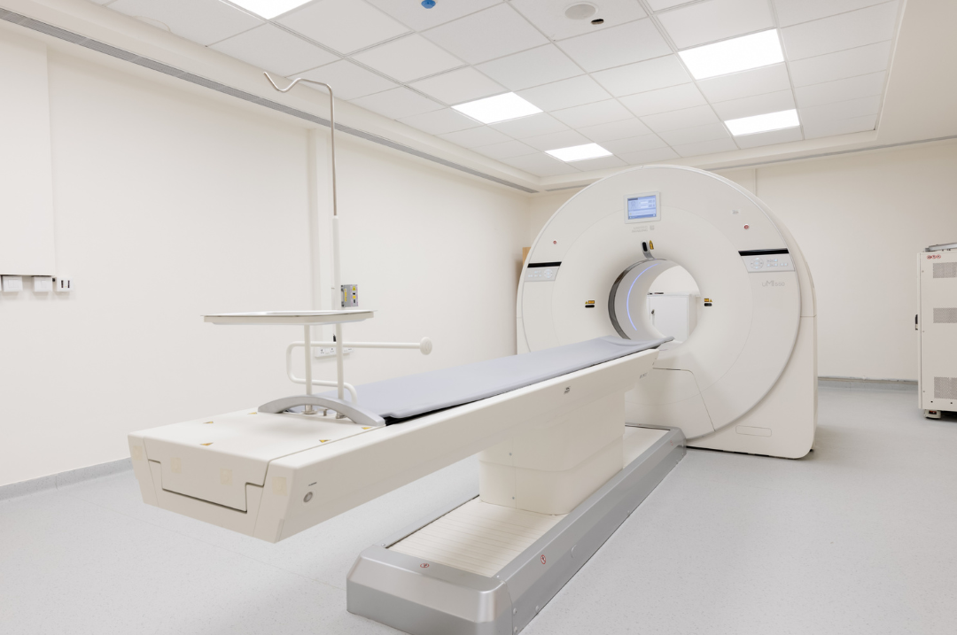
On the day of your scan, you will be asked to lie down on a table in the imaging room. A healthcare provider will explain the procedure and ensure you're ready for the scan.
A small amount of the radioactive CXCR4-specific tracer will be injected into a vein, typically in your arm. This injection is generally painless, though you might feel a slight pinch.
After the injection, you will need to wait for about 45-60 minutes. This gives the tracer time to circulate through your body and bind to the CXCR4 receptors in the tissues
During the scan, you will lie still on the table while the PET and CT scanners take images of your body. The PET scan will provide information about the biological activity, while the CT scan gives detailed anatomical images. The scanning process usually takes around 10 minutes, depending on the area being scanned.
Once the scan is complete, you can resume normal activities. The radioactive tracer used in the procedure will be naturally eliminated from your body over time, mainly through urination.
Benefits of a CXCR4 PET-CT Scan:
-
Early Detection of Multiple Myeloma
The scan can help detect bone marrow involvement and extramedullary plasmacytomas, which are often seen in multiple myeloma.
-
Accurate Tumor Staging
By providing both functional (PET) and anatomical (CT) images, the CXCR4 PET-CT scan allows for more accurate staging of multiple myeloma, aiding in treatment planning.
-
Treatment Monitoring
It helps doctors monitor how well multiple myeloma treatments are working by assessing changes in CXCR4 expression.
-
Identifying Adrenal Tumors
The scan can help evaluate adrenal adenomas, assisting in differentiating functional and non functional adenoma’s.
Subsribe To Our Newsletter
Stay in touch with us to get latest news and special offers.