Advanced Imaging for Precise Kidney Cancer Detection
CAIX Digital PET/CT Scan
A targeted PET/CT scan targeting CAIX receptors to accurately detect and assess
renal cell carcinoma (kidney cancer) lesions.
About CAIX Digital PET/CT Scan
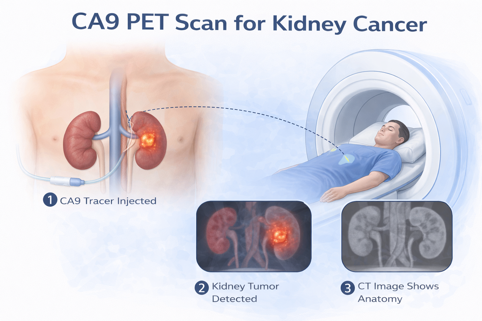
A CAIX PET/CT scan is an advanced imaging technique used to diagnose, stage, and plan treatment for renal cell carcinoma (RCC). It combines PET and CT to provide both functional and anatomical detail. A CAIX-targeted tracer is injected and binds to CAIX, a protein highly expressed in clear cell RCC. PET highlights cancer activity, while CT shows precise anatomy. Together, they enable accurate detection, localization, and assessment of tumor spread, supporting better treatment planning.
Why is a CAIX PET/CT Scan Performed?
CAIX PET/CT scans play an important role in the evaluation and management of kidney cancer, particularly clear cell renal cell carcinoma. Key clinical uses include:
01. Detection of Kidney Cancer
CAIX PET/CT scans are highly sensitive in identifying primary renal tumors, especially clear cell renal cell carcinoma. They help detect lesions within the kidney
with greater molecular specificity.
02. Staging of Renal Cell Carcinoma
The scan helps determine whether the cancer has spread beyond the kidney to lymph nodes, bones, lungs, or other organs, providing essential information for
accurate cancer staging.
03. Detection of Metastatic Disease
CAIX PET/CT is valuable for identifying small metastatic lesions that may not be easily visible on conventional CT or MRI scans.
04. Evaluating Treatment Response
The scan can be used to assess how well kidney cancer is responding to targeted therapy, immunotherapy, or systemic treatments, helping physicians adjust treatment strategies when needed.
05. Planning Targeted Therapies
By precisely identifying the location and extent of disease, CAIX PET/CT helps guide surgical planning, radiation therapy, and emerging molecular targeted
therapies for renal cell carcinoma.
How is the CAIX PET/CT Scan Performed?
FDG PET/CT scans are used in many important areas of healthcare, and they can help doctors make better decisions for treating a variety of conditions:
-
Preparation
Patients may be advised to avoid heavy meals for 2–3 hours before the scan. A recent serum creatinine report may be required if contrast CT imaging is planned.
-
Injection of CAIX Tracer
A small amount of radioactive tracer that binds to CAIX receptors is injected into a vein. The tracer selectively attaches to kidney cancer cells that express CAIX.
-
Resting Period
After the injection, patients rest for approximately 45–60 minutes to allow the tracer to circulate and accumulate in CAIX-expressing cancer cells.
-
The Scanning Process
During the scan, the patient lies on a table that moves slowly through the PET/CT scanner. It is important to remain still during the scan to obtain clear images. The scanning process under the scanner typically takes around 10 minutes.
-
Post-Scan
After completion of the scan, patients can resume normal daily activities. Drinking fluids may be recommended to help eliminate the tracer from the body.
Subsribe To Our Newsletter
Stay in touch with us to get latest news and special offers.